
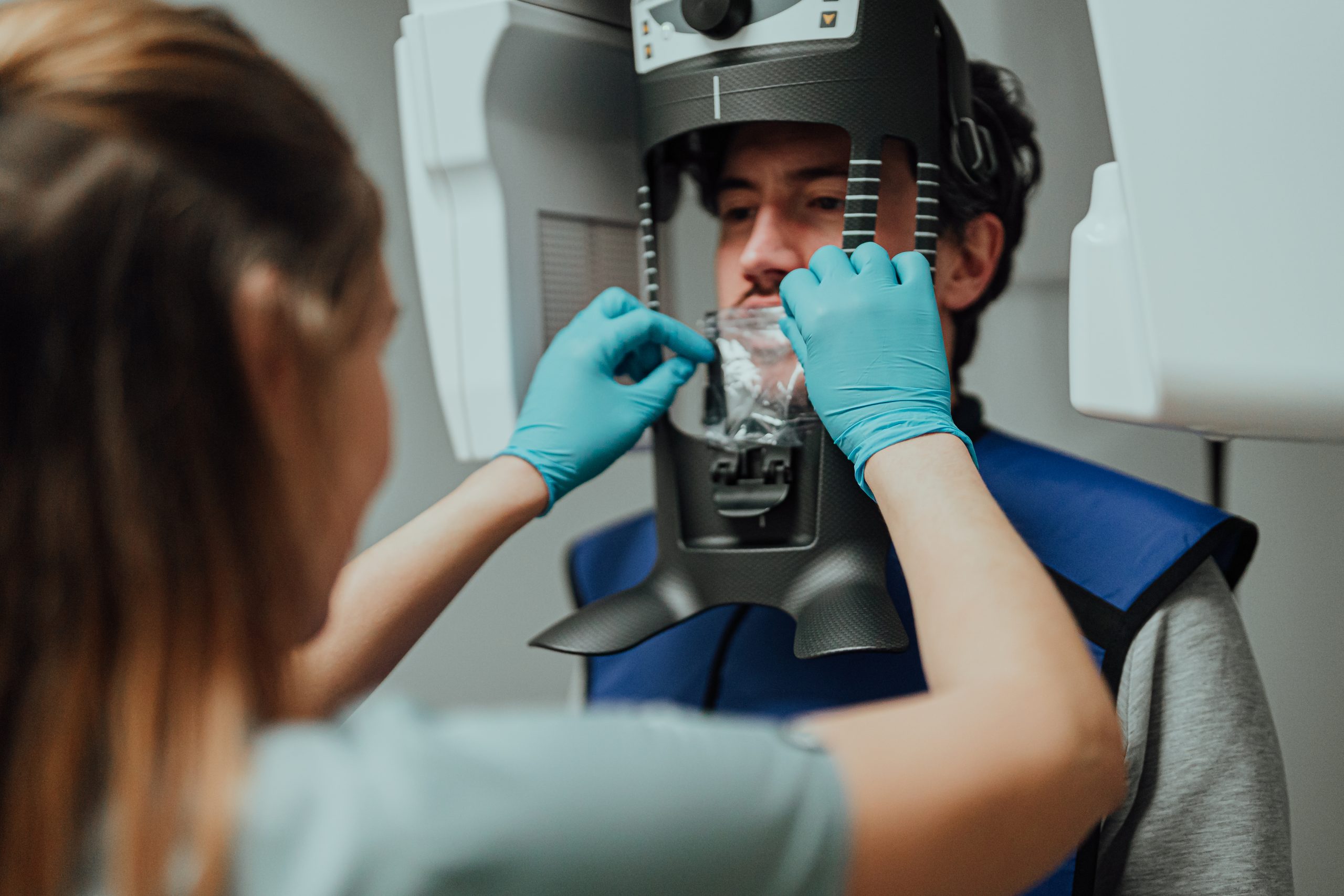
3D tomography
2000+
Happy smiles per month
97%
Client satisfaction
60+
Team members
25000+
Amazing clients
Why and when it is needed?
In dentistry 3D visualisation expands the diagnostic opportunities and enables to have a look at the entire mouth cavity not only one tooth. This method of equipment diagnostics is widely used in orthodontics, face and jaw surgery, implantology and plastic surgery. 3D dental image is necessary:
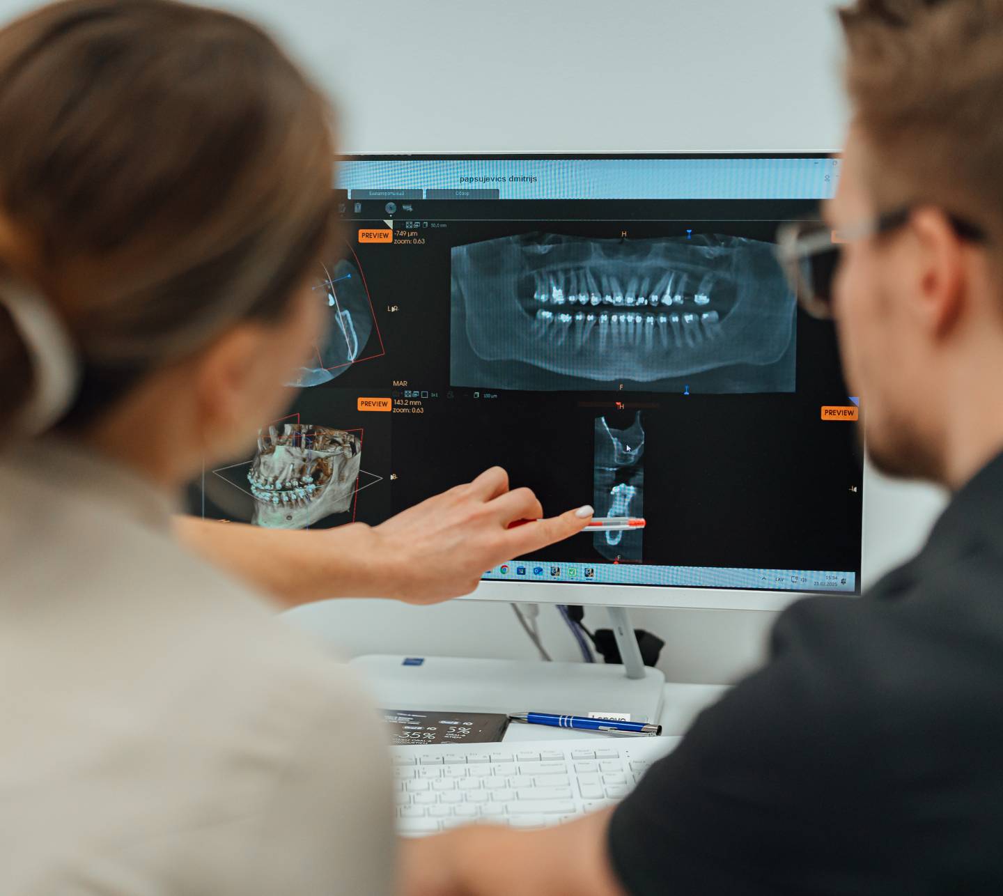
Types of X-rays
- Provides a full overview of the condition of teeth, jaws, and surrounding structures.
- Is used for detailed assessment of a specific tooth or area.
- This technology delivers volumetric imaging of the jaw and face area for precise diagnostics and treatment planning.
- Helps analyze facial symmetry, bite alignment, and supports orthodontic treatment planning.
Sign up for a consultation
Planning a visit to the dentist, but don’t know where to start? Book a consultation before treatment and get answers to all your questions. We will help you understand the necessary procedures and take the first step to a healthy smile!

What our patients say
Positive feedback from patients confirms the high level of trust in our work. We greatly value every opinion, as it helps us continuously improve the quality of our services.
F.A.Q.
3D X-ray imaging is a method that uses X-ray radiation to create a three-dimensional image of the teeth, jaws, and surrounding tissues. It provides detailed layers of information, allowing dentists to better visualize the anatomical structures.
3D X-rays are recommended for planning implant placement, in orthodontics, for assessing complex root canal cases, or when there are anomalies that require more detailed investigation. During check-up diagnostics, a 3D X-ray is also performed for a more accurate assessment of the condition of the teeth and gums.
Yes, 3D X-ray is a safe procedure as it uses a minimal amount of radiation. Advanced technology allows for reduced exposure compared to traditional X-ray images.
The 3D X-ray procedure takes just a few minutes. After the scanning is complete, the images are immediately available for analysis by the dentist.
Yes, a 3D X-ray can be performed even if you have dental prosthetics or implants. It provides useful information about the condition of the surrounding tissues and allows dentists to plan further treatment.
3D X-ray imaging allows for more detailed and precise images, aiding in diagnosis and treatment planning. This reduces the likelihood of errors and improves treatment outcomes.
Sign up for a consultation
Planning a visit to the dentist, but don’t know where to start? Book a consultation before treatment and get answers to all your questions. We will help you understand the necessary procedures and take the first step to a healthy smile!